This article looks at specific accusations in the citizens petition supplement attachment 1
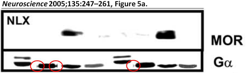
Example #1: “Manipulated WB”. The CP claims that different WBs were “spliced together” to make a figure in a 2005 paper. They point out a “bowtie” effect of several bands as supporting evidence. However, anyone with WB experience has seen this bowtie effect. In theory, it can be caused by proteins sticking to the sides of the wells or a small amount of extract entering the space between the wells and the plates that support the gel on each side. This effect is often seen when the top of the gel is exposed to air too long prior to loading.
Example #2: “Falsified WB”. This claim states that the same beta-actin control WB was used in 2 different papers published 5 years apart. The CP takes a beta-actin WB, which is low resolution, stretches it vertically, then blows it up and compares it to a WB published 5 years earlier. Although they claim the blots are identical, to me they look completely different. For example: 1) the left side of the band in lane 1 of the 2005 publication has a notch that is not in the 2010 WB; 2) band images between lanes 2 and 3 are clearly different; 3) the right sides of the bands in lane 4 are also different. I also have difficulty taking this claim seriously. The labs likely ran many beta-actin WBs between the 5-year timeframe the 2 papers were published, but it is claimed they somehow elected to risk their scientific careers to include 4 control lanes from a previous publication? A new beta-actin WB could be run in half a day. This accusation suggests that the scientists who contributed to the CP are not very experienced in molecular biology.

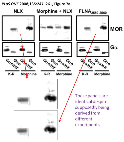
Example #3: “Reused WB”. This claim takes 2 WB panels of a figure and compares them side-by-side, suggesting they are identical. This claim could have some credibility, though it is more likely a mistake in constructing the figure for publication than scientific misconduct. Although I can’t say for sure that the 2 WBs in question are indeed the same, they do look very similar, though one is a slightly darker exposure. When WBs are exposed to film, we generally get several exposures from light to dark. If the films aren’t labelled properly, things can get mixed up when the graduate student/post-doc generates the figure for publication, especially if two of the WBs look similar. If the 2 blots are indeed the same, the PI, other authors, or reviewers probably should have asked if this was indeed an error. I’m inclined to think this was an error in making the figure and not intentional since there is nothing covert going on here- the 2 WBs are basically side-by-side in the same figure.
Example #4: “Band insertion into WBs”. The concerns raised here include: 1) irregular spacing between lanes; 2) FLNA bands not looking correct since it is a large (290 kDa) protein; 3) bands looking identical between lanes; and 4) white halos around bands. In my opinion, this claim is completely baseless and shows a lack of experience of the scientists that generated the CP. Irregular spacing between bands is routinely seen with self-poured gels. It is explained by the teeth of the comb used to cast the gel not being completely straight when the gel is polymerized. The claim that FLNA shouldn’t run very far in a gel because of its large size is inaccurate because proteins will run as far through a gel as a current is applied. Longer gels could have been used. Also, the researchers could have used gradient gels that allow for larger proteins to be better resolved in the gel. As far as the control beta-actin bands looking identical, they are basing this assumption on a common “tadpole-like” appearance of the bands. However, this tadpole effect is commonly seen if the gel is polymerized next to an air current (e.g. in a fume hood). The air current can cause the sides of the wells to lose moisture during polymerization resulting in a tadpole-like effect on the same sides of the wells of each lane, which is seen here. As far as halos around bands, most films used to expose WBs have a threshold level of exposure that is designed to limit background. You often see this halo effect directly on the exposed film, especially with stronger bands. The white halo could also be a compression effect, caused when the figure levels were adjusted for publication.

Concern 3.2 “Evidence of data manipulation from human tissue”. It is claimed that Figure 12 of a 2017 paper shows a WB with 12 lanes for the control protein NR1 and 13 lanes for PCLgamma1. It is also suggested that WBs from different experiments were spliced together to make the figure. The 12/13 lane issue is an obvious error that likely occurred when making the figure for publication, but in no way rises to the level of scientific misconduct. As far as splicing together 2 different WBs, please refer to Response to General Concern #1 above about manipulating low-resolution figures and spliced WBs. I honestly don’t see much evidence of splicing for the top WB. Also, on the bottom WB, the change in pixels appears to wrap around the top of the band on the left side of the proposed splice site. It would be difficult to crop the figure this way. Another thing to point out is that the 3 lanes on the right of both WBs are running lower in the gel than the band directly to the left, which is slightly higher than the other bands to its left. However, the proposed splice sites are 4 lanes from the right on the top WB and 3 lanes from the right on the bottom WB. These factors support that the figure was not generated by splicing 2 WBs spliced together.

Appendix items/Additional areas of concern:
Claim #4: It is claimed that the top WBs cannot be FLNA based on the banding pattern. Please see Response to Example #4 above. Also, note that Albumin was used as the control protein here, which is a comparatively large (66 kDa) for a control protein and suitable for analysis of large proteins, such as FLNA.

Claim #6: It is claimed that several papers between 2010-2017 don’t show control WBs for co-immunoprecipitation experiments that evaluate the interaction of beta-amyloid with alpha7-nicotinic acetylcholine receptors. This claim is true, but there is a very good reason for it. Beta-amyloid are small (10 kDa) peptides of 40-43 amino acids in length. Their small size makes them very difficult to analyze by WB, other than running specialized gels that would likely preclude the analysis of larger interacting proteins, such as alpha7nAChR (60+ kDa). Most companies pre-validate their antibodies for use in certain applications, such as immunoprecipitation experiments.

E.2 Additional suspicious WBs (1) and probable band duplication (2):
1) Suspicious WBs. The CP shows several examples (pages 30-31) of control bands looking the same between WBs suggesting that the researchers must have duplicated these images. To me, this is a ridiculous claim. I see clear differences between the bands in question and I think most others do as well.
2A) Probable band duplication. Regarding the white halo around bands on WBs, see Response to Example #4 above.
2B) FLNA bands look identical (page 33). The CP highlights 3 FLNA bands that they claim are duplicated, but I see clear differences. For example, differences are seen with the bottom of the right and middle bands and top of the right bands.
2C) Five IRbeta bands look identical (page 34). There are clear differences in the sizes of the notch at the bottom left of each band, as well as the slopes of the bands on the top right.
2D) Clipping multiple blots together (page 35). The clip effect that is seen when the proteins run through an air bubble in the gel, which is common with self-poured gels.
2E) Tampering of WBs (pages 37-39). It is claimed that bands are inserted into various WBs in a 2006 Nature Medicine publication. This claim is borderline ridiculous and is likely why it appears at the end of the CP. Dr. Wang is first author on this publication, so the implication being made is that he started manipulating data early in his career and continues to do so today as head of an academic laboratory. Firstly, Nature Medicine is an outstanding journal, so the data was thoroughly vetted by experts in the field prior to publication. See Response to General Concern #1 regarding manipulation of low-resolution WB images and drawing conclusions. Also, several of the examples shown as evidence of data manipulation support the opposite. For example, on page 38 the background pixels around the bands are irregularly shaped. It is hard to believe that Dr. Wang would have cropped each of the bands for insertion as irregular shapes (which would be difficult in 2006) instead of just using a box shape. Also, if differences in background pixels are evidence of data manipulation, why are there also irregular shaped pixels around the text in the figures? Were they copied from other figures as well? It is obvious that the differences in background pixels observed are the direct result of the scientists who constructed the CP altering the contrast and brightness of already low-resolution figures.

In the scope of things these arguments are scientifically meaningless.. Besides, Would you want to spend your life being an expert on such blots and protein migrations patterns on gels? Besides all this, i The real issue is that Wangs lab work was reconfirmed by Quanterix (SP?). Thus, two independent labs, using different methodologies and different samples (plasma vs cerebrospinal fluid) confirmed each other. It does not get any more credible than that.
LikeLiked by 2 people
Don’t forget that when Quanterix came out with the statement saying they did not interpret the data and the stock sold off. Its a lab, not a university, they don’t interpret the data for you…
LikeLike
Not only the above but SAVAs unquestioned results on Cognitive Improvement. Of pertinence is a recent piece from Scientific American should bode well for SAVA: And, I think this is something all of us expect from the FDA Advisory panel in regard to SAVA in its upcoming Phase 3 Trials.
The FDA approves an increasing number of drugs, including the controversial Alzheimer’s drug aducanumab, based on changes in surrogate endpoints, measurements such as blood tests that substitute for clinical outcomes and constitute lower-quality evidence than direct observations. If surrogates for clinical improvement are good enough for the government to establish clinical efficacy, then direct observations of clinical benefit, a much higher bar, should suffice to establish currently accepted medical use.
LikeLiked by 2 people
I enjoy your work greatly.
I will make sure http://www.sava-ad.com links to your great work here!
LikeLiked by 1 person